41 dissected cow eye labeled
Cow Eye Dissection - Perkins School for the Blind Step-by-step instructions for a science lab to dissect a cow eye. This is a great activity for middle and high school students. It allows them to participate in a dissection, while teaching the anatomy and structure of the eye. To get started, watch the YouTube video Cow's Eye Dissection: Exploratorium. It can be a support for guiding ... Cow Eye Dissection & Parts of the Eye Diagram | Quizlet White, outermost layer of the eye. Helps maintain shape and gives attachment to muscles. photoreceptors The cells in the retina that respond to light (rods and cones) rods Photoreceptor cells in the eye that detect black, white, and gray cones Photoreceptor cells in the eye that detect color aqueous humor
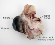
Cow Eye Dissection & Anatomy Project | HST Learning Center On the posterior side of the eye, nestled in the fat and muscle tissue, there is a noticeably round protuberance that feels stiffer than the surrounding tissue. This is the optic nerve, and it sends the images collected in the eye to the brain. Cow Eye Dissection: Internal Anatomy. 1. Place the cow's eye on a dissecting tray. The eye most ...
Dissected cow eye labeled
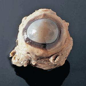
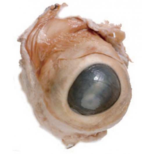
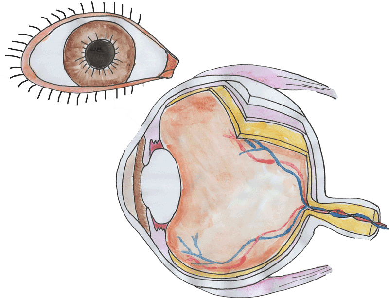
Cow's Eye Dissection | Exploratorium Learn how to dissect a cow's eye in your classroom. This resource includes: a step-by-step, hints and tips, a cow eye primer, and a glossary of terms. Cow Eye Dissection - Biology LibreTexts 1. Examine the outside of the eye. You should be able to find the sclera, or the whites of the eye. This tough, outer covering of the eyeball has fat and muscle attached to it. 2. Locate the covering over the front of the eye, the cornea. When the cow was alive, the cornea was clear. In your cow's eye, the cornea may be cloudy or blue in color. 13.7: Cow Eye Dissection - Biology LibreTexts 1. Examine the outside of the eye. You should be able to find the sclera, or the whites of the eye. This tough, outer covering of the eyeball has fat and muscle attached to it 2. Locate the covering over the front of the eye, the cornea. When the cow was alive, the cornea was clear. In your cow's eye, the cornea may be cloudy or blue in color. 2.
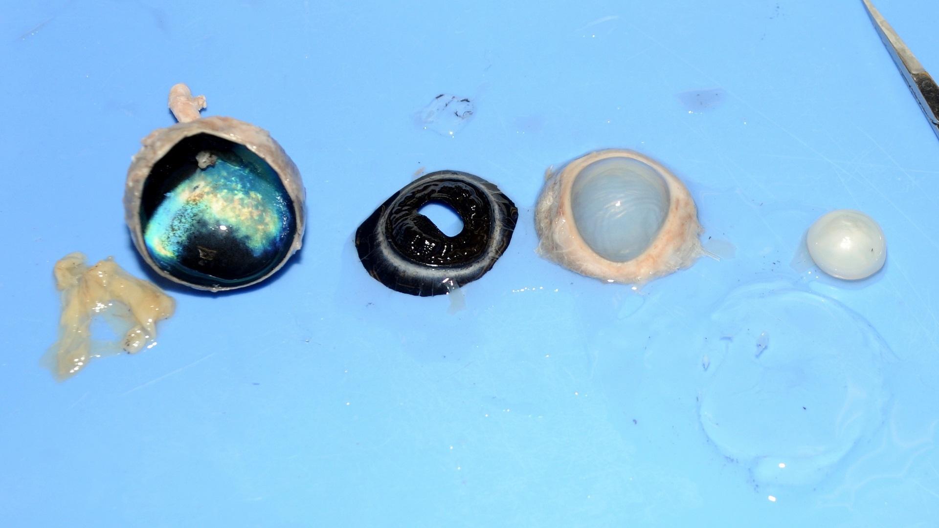
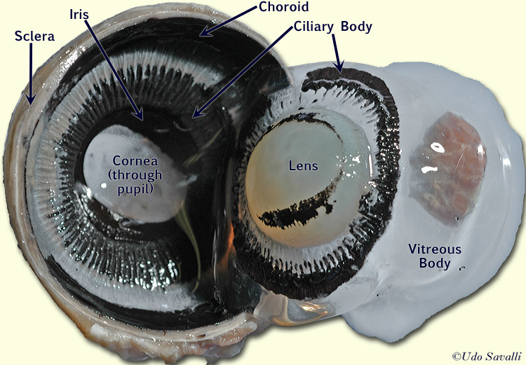
Dissected cow eye labeled. 13.7: Cow Eye Dissection - Medicine LibreTexts 1. Examine the outside of the eye. You should be able to find the sclera, or the whites of the eye. This tough, outer covering of the eyeball has fat and muscle attached to it 2. Locate the covering over the front of the eye, the cornea. When the cow was alive, the cornea was clear. In your cow's eye, the cornea may be cloudy or blue in color. 2. Cow Eye Dissection Kit for Kids Animal Anatomy Labs | HST Inside this cow eye dissection kit, you'll find: a preserved cow eye, a full-color photographic eye dissection guide, and the dissection tools/lab equipment you'll need: a #22 broad-blade scalpel, scissors, and a sturdy disposable dissecting tray. You'll get see how a cow's eye is like a human eye as you closely investigate parts from the iris ... Cow Eye Dissection Guide - Google Slides Dissection 101: Cow Eye Use the point of a scissors or a scalpel to make an incision through the layers of the eye capsule (similar to figure 1); there are three layers from the exterior:... Cow's Eye Dissection | Exploratorium Step 1: The cow's eye Here's a cow's eye from the meat company. The white part is the sclera, the outer covering of the eyeball. The blue is the cornea, which starts out clear but becomes cloudy after death. Step 2: Muscles move the eye Without moving your head, look up. Look down. Look all around.
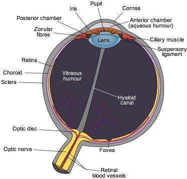
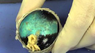
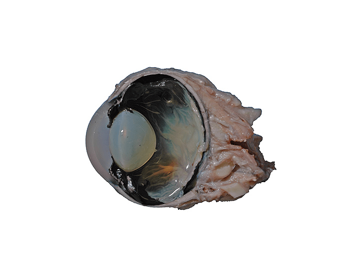
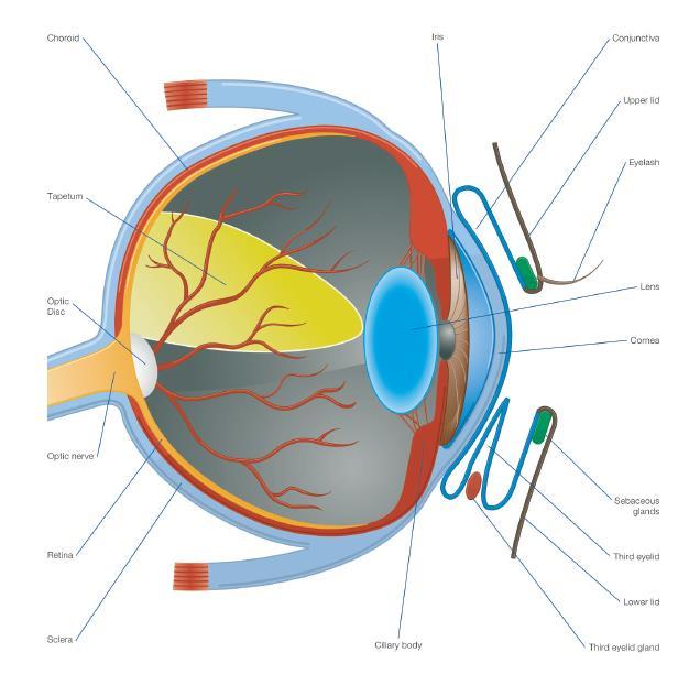
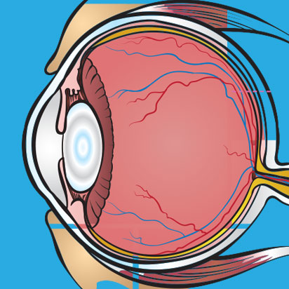
Cow's Eye Dissection - Eye diagram - Exploratorium Learn how to dissect a cow's eye in your classroom. This resource includes: a step-by-step, hints and tips, a cow eye primer, and a glossary of terms. Cow's Eye Dissection - Eye diagram PDF COW'S EYE dissection - Exploratorium A cow's iris is brown. Human irises come in many colors, including brown, blue, green, and gray. lens clear, flexible structure that makes an image on the eye's retina. The lens is flexible so that it can change shape, focusing on objects that are close up and objects that are far away. myelin The fatty layer that surrounds each nerve fiber. Cow Eye Dissection | Carolina.com Cow Eye Internal Anatomy Hold the eye between your thumb and forefinger, as shown below. Using scissors or a scalpel, carefully cut the eye in half, separating the front and back of the eye. Examine the inside front portion of the eye. Remove the gelatinous vitreous humor and hard lens. PDF Home Science Tools Resource Center Home Science Tools Resource Center
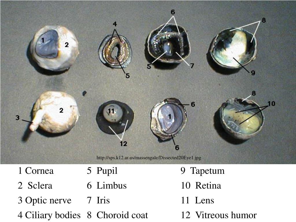
Solved 1. Identify the labeled structures in the | Chegg.com Identify the labeled structures in the accompanying photographs of a dissected cow eye. a. Sclera b. Cornea c. choroid? d. Optic Disc - Retina UNIT 17 | General and Special Senses 395 Show transcribed image text Expert Answer 1st step All steps Final answer Step 1/5 Label a Sclera Cow's Eye Dissection - step 1 - Exploratorium At the Exploratorium, we dissect cows' eyes to show people how an eye works. This Web site shows photos and videos of a dissection. If you try this at home, wash your hands after the dissection. Wear latex gloves if you have cuts in your hands. Here's a cow's eye from the meat company. Cow Eye Dissection - Carolina Knowledge Center Carolina's Perfect Solution® cow eye dissection introduces students to the anatomy of the mammalian eye. This activity allows students to identify the major structures of the eye. The activity supports 3-dimensional learning and builds toward the following: NGSS Scientific and Engineering Practice: Developing and Using Models. Cow Eye Dissection - The Biology Corner 1. Examine the outside of the eye. You should be able to find the sclera, or the whites of the eye. This tough, outer covering of the eyeball has fat and muscle attached to it. 2. Locate the covering over the front of the eye, the cornea. When the cow was alive, the cornea was clear. In your cow's eye, the cornea may be cloudy or blue in color.
Preserved Cow Eyes
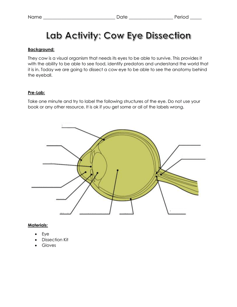
PDF Cow Eye Dissection Guide DISSECTION OF THE COW EYE Please make sure to wear gloves and safety glasses when you are dissecting, and make sure to clean up thoroughly after the lab. Also, the cow eyes can be rather slippery, so use caution when handling and cutting them. You will need a scalpel and forceps. 1. First, identify the most external structures of the eye.

What an eye is made up of | Cow eyes, Dissection, How to ...
Solved Name the dissected cow eye structure labeled #6.A) - Chegg Name the dissected cow eye structure labeled #6. A) Ciliary body B)Lens C) Vitreous humor D) Iris E)Pupil Expert Answer 100% (1 rating) 1st step All steps Final answer Step 1/2 Name the dissected cow eye structure labeled. 1. Cornea. View the full answer Step 2/2 Final answer Previous question Next question This problem has been solved!
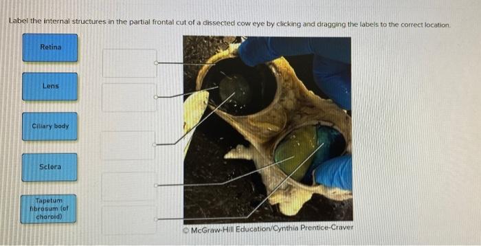
Solved Label the internal structures in the partial frontal ...
13.7: Cow Eye Dissection - Biology LibreTexts 1. Examine the outside of the eye. You should be able to find the sclera, or the whites of the eye. This tough, outer covering of the eyeball has fat and muscle attached to it 2. Locate the covering over the front of the eye, the cornea. When the cow was alive, the cornea was clear. In your cow's eye, the cornea may be cloudy or blue in color. 2.
Dissection 101 | Detailed Cow Eye Dissection Video (Part 2 of 2)
Cow Eye Dissection - Biology LibreTexts 1. Examine the outside of the eye. You should be able to find the sclera, or the whites of the eye. This tough, outer covering of the eyeball has fat and muscle attached to it. 2. Locate the covering over the front of the eye, the cornea. When the cow was alive, the cornea was clear. In your cow's eye, the cornea may be cloudy or blue in color.

Cow eye - internal anatomy | Cow eyes, Medical anatomy, Anatomy
Cow's Eye Dissection | Exploratorium Learn how to dissect a cow's eye in your classroom. This resource includes: a step-by-step, hints and tips, a cow eye primer, and a glossary of terms.
NEUR 320: Art and Vision
Eye Dissection

7,161 Human Eye Diagram Images, Stock Photos & Vectors ...
Cow Eye Dissection & Anatomy Project | HST Learning Center

Diagram of human eye anatomy with label 1868583 Vector Art at ...

Fresh cow eye dissection (Part 2): Front of eye
Human eye diagram - Eye Anatomy - Online Biology Dictionary
Cow Eye Dissection

Eye Anatomy

Science - *Zakiabizk*
Cow Eye Dissection Pre-lab Key

US HISTORY101 - Cow Eye Dissection Post Lab - Aidan - Eye ...
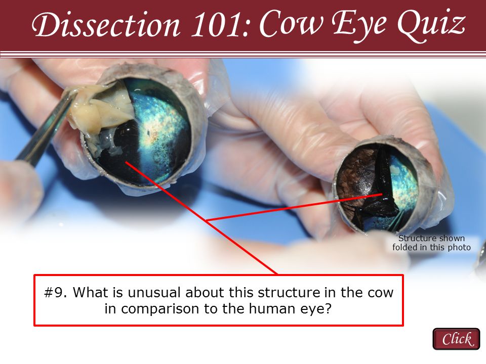
Cow Eye Quiz Dissection 101: Click - ppt video online download

Cow Eye Dissection Pre-lab.docx - Cow Eye Dissection Pre-Lab ...

13.7: Cow Eye Dissection - Biology LibreTexts
Cow Eye, Preserved, Bulk Bag

Solved Name the dissected cow eye structure labeled #6.A ...

Cow Eye Dissection Kit
Cow Eye Labeling Quiz
PPT - COW EYE DISSECTION PowerPoint Presentation, free ...
Cow's Eye Dissection - Eye diagram
BIO201-Cow Eye
Anatomy of the eye

Biology Lab 10 Cow Eye Dissection Diagram | Quizlet
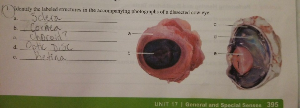
Solved 1. Identify the labeled structures in the | Chegg.com
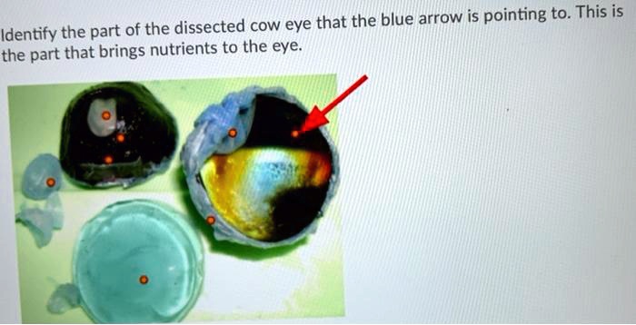
SOLVED: Identify the part of the dissected cow eye that the ...
Eyes - Layers of Learning

Detailed Cow Eye Dissection: Part II (Jr. High, High School and College Review)

"Carolina® Eye Dissection Mat"
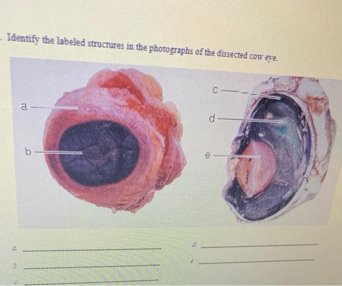
Solved Identify the labeled structures in the photographs of ...

Dissected cow eye Diagram | Quizlet

Lab 3- Eye Surgeries

Cow Eye Dissection - Perkins School for the Blind

Young Scientist's Eye Dissection Kit: Science Lab Biology ...

Eye Dissection
Cow Eye Dissection | Carolina.com
Post a Comment for "41 dissected cow eye labeled"