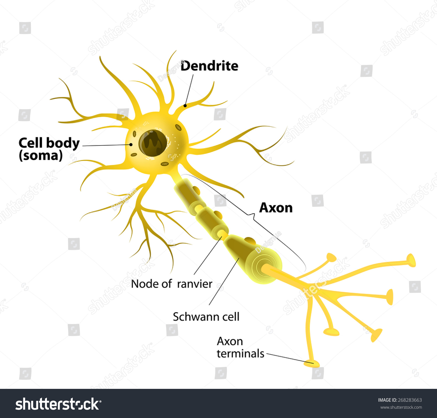
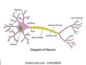
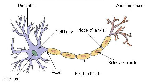
40 neuron diagram labeled
Neuron Diagram & Types | Ask A Biologist - Arizona State University Unipolar neurons are also known as sensory neurons. They have one axon and one dendrite branching off in opposite directions from the cell body. These cells pass signals from the outside of your body, such as touch, along to the central nervous system. Bipolar neurons have one axon and only one dendrite branch. Labeled Neuron Diagram Pictures, Images and Stock Photos Search from Labeled Neuron Diagram stock photos, pictures and royalty-free images from iStock. Find high-quality stock photos that you won't find anywhere else.
What is Perceptron: A Beginners Guide for Perceptron Aug 11, 2022 · A neural network link that contains computations to track features and uses Artificial Intelligence in the input data is known as Perceptron. This neural links to the artificial neurons using simple logic gates with binary outputs. An artificial neuron invokes the mathematical function and has node, input, weights, and output equivalent to the cell nucleus, …
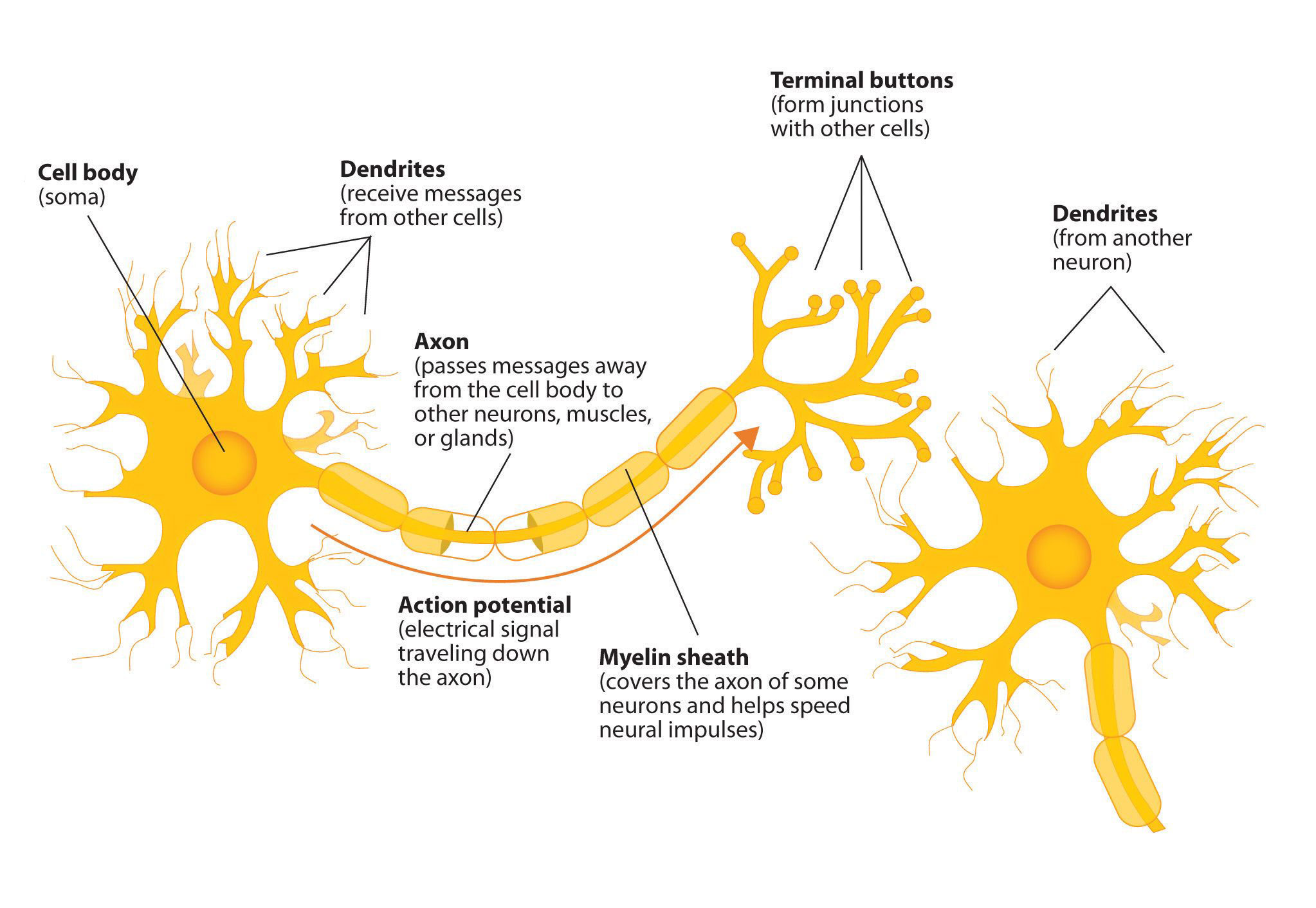
Neuron diagram labeled
Neuron Diagram Labeled Pictures, Images and Stock Photos Search from Neuron Diagram Labeled stock photos, pictures and royalty-free images from iStock. Find high-quality stock photos that you won't find anywhere else. Video, Trending searches, Basketball, Fire, Texture, Chinese new year, Black history, Football, Popular categories, Lunar New Year Videos, Aerial videos, Abstract videos, Label Neuron Anatomy Printout - EnchantedLearning.com Read the definitions, then label the neuron diagram below. axon - the long extension of a neuron that carries nerve impulses away from the body of the cell. cell body - the cell body of the neuron; it contains the nucleus (also called the soma) dendrites - the branching structure of a neuron that receives messages (attached to the cell body) Diagram Quiz on Neuron Structure and Function (Labeling Quiz) This labelled diagram quiz on Neuron is designed to assess your basic knowledge in Structure and Function of Neuron. Choose the best answer from the four options given. When you've finished answering as many of the questions as you can, scroll down to the bottom of the page and check your answers by clicking ' Score'.
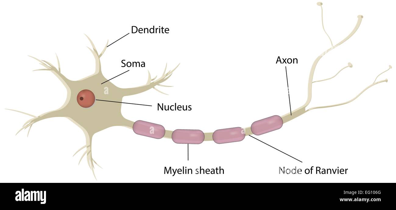
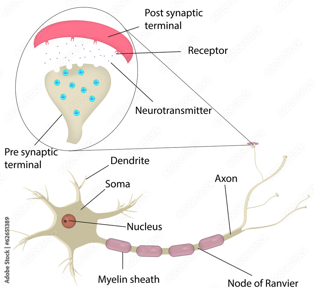
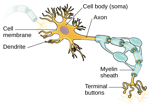
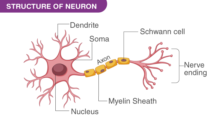
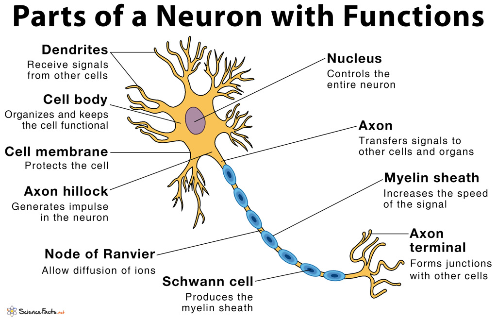
Neuron diagram labeled. neuron model labeled Diagram | Quizlet Start studying neuron model labeled. Learn vocabulary, terms, and more with flashcards, games, and other study tools. Single-neuron projectome of mouse prefrontal cortex - Nature Mar 31, 2022 · Top, a diagram and an example neuron with the over-represented pattern of projections to MOp, SSp, and SSs. Extended Data Fig. 6 Diversity of projections to lateral and central cortical subnetworks. Labeled Neuron Diagram| EdrawMax Template The following labeled diagram shows the parts of a neuron. In order to make it more understandable to the students, we have added all the functions of the Neuron in the labeled diagram. The major parts of the Neuron are Dendrites, Cell Body, Cell Membrane, Axon Hillock, Node of Ranvier, Schwann Cell, Axon Terminal, Myelin Sheath, Axon, and Nucleus. Neurons (With Diagram) - Biology Discussion A neuron is a structural and functional unit of the neural tissue and hence the neural system. Certain neurons may almost equal the length of body itself. Thus neurons with longer processes (projections) are the longest cells in the body. Human neural system has about 100 billion neurons. Majority of the neurons occur in the brain.
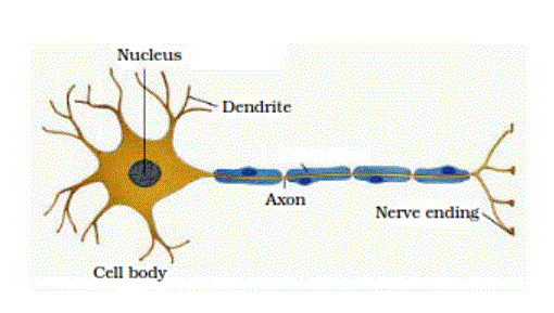
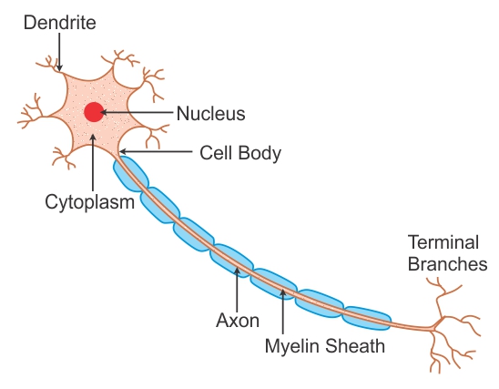
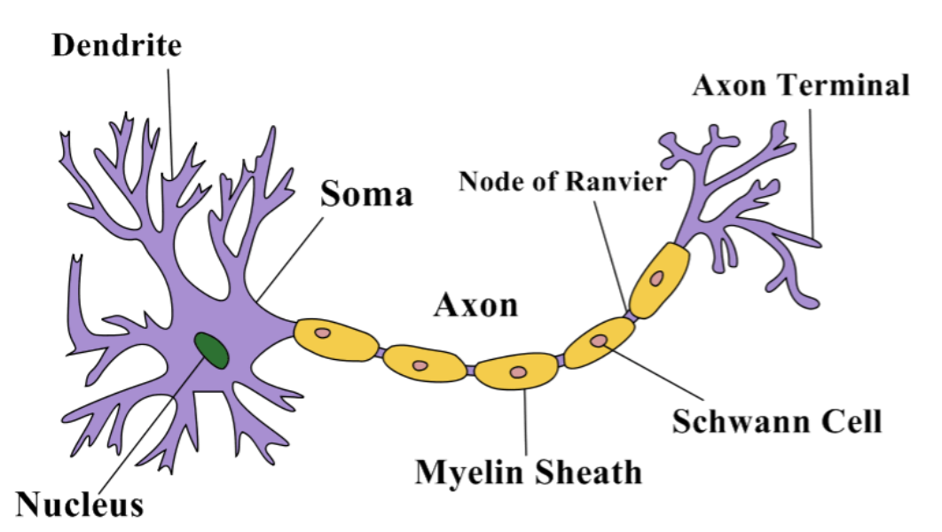
Neuron Diagram Labeled | EdrawMax Template It is an effective form of self-assessment, enabling students to check their understanding. In the following diagram, we have illustrated the important parts of the Neuron. In the following Neuron labeled diagram, we have dendrite, cell body, axon, myelin sheath, Schwann cell, a node of Ranvier, axon terminal, and nucleus. Gate control theory - Wikipedia The same neurons may also form synapses with an inhibitory interneuron that also synapses on the projection neuron, reducing the chance that the latter will fire and transmit pain stimuli to the brain (image on the right). ... The authors had drawn … Templates Community | EdrawMax Discover, learn, and be inspired by the top EdrawMax visuals, curated by our users. Then join them by sharing your own! Neuron under Microscope with Labeled Diagram - AnatomyLearner The neuron structure has two main components: the cell body and the neuron processes (axons and dendrite). Let's see the neuron histology slide labelled diagram and try to find out the below-mentioned characteristics -, Presence of an identifiable cell body (soma) that locates in the brain's grey matter (according to the slide image).
how to draw structure of neuron/neuron diagram labelled ... - YouTube Please watch: "cell structure and functions / animal cell vs plant cell / parts of cell / ch 8 science class 8 cbse" ... A Guide to Understand Neuron with Neuron Diagram The students can find the neuron labeled diagram in this option. Step 3: Once the students have selected their diagrams, they can smoothly work on them. They can modify the diagram according to their choice. It can help them to get a high-quality neuron labeled diagram apt for their projects and dissertation papers. Neuron Label Teaching Resources | Teachers Pay Teachers NERVE CELLS AND SYNAPSES DIAGRAM WORKSHEETIncluded in this resource:Nerve Cell Diagram Worksheet - Looking at parts of the nerve cell, students can label and describe the functions (e.g. nucleus, axon, dendrites, myelin sheath etc). Students are also asked to define what a neuron is and the three types or neuron, linked to a simple diagram. 7. Artificial neural networks - Massachusetts Institute of … Neuron Unit Synapse Connection Synaptic strength Weight Firing frequency Signals pass fromUnit output Table 1 (left): Corresponding terms from biological and artificial neural networks. Adapted from Adapted from Mehrotra, Mohan, & Ranka. Figure 1 (below): Schematic diagram of a standard neural network design. the input units
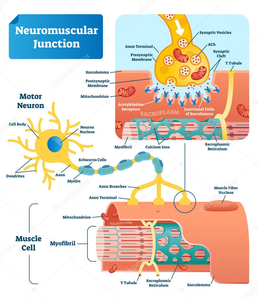
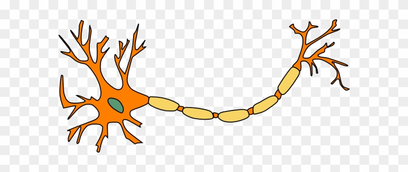
Labeled Neuron Diagram | Science Trends Labeled Neuron Diagram, Alex Bolano, 29, May 2019 | Last Updated: 3, March 2020, Neurons are the basic organizational units of the brain and nervous system. Neurons form the bulk of all nervous tissue and are what allow nervous tissue to conduct electrical signals that allow parts of the body to communicate with each other.
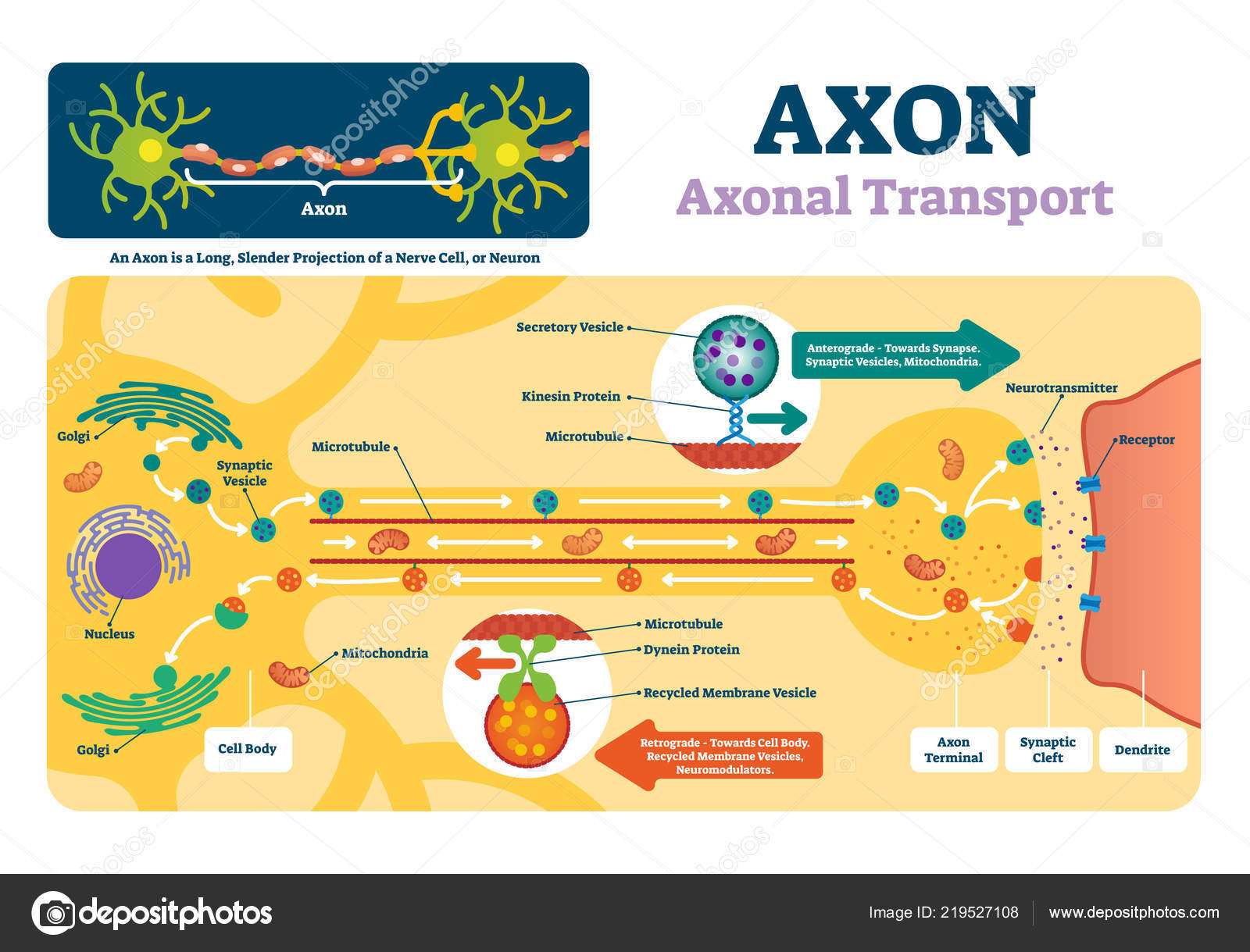
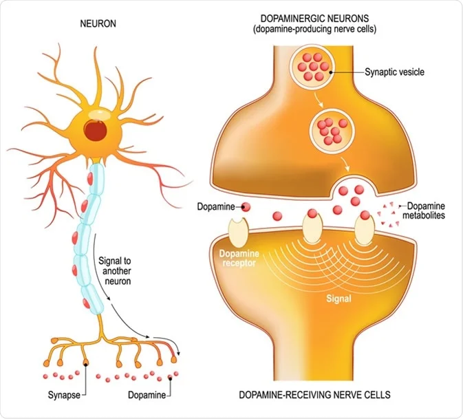
Nodes of Ranvier: Location And Function (With a Labeled Diagram ... A neuron receives a signal from another neuron in the form of chemical neurotransmitters. These are produced by the terminals of the conducting neuron and are received by the dendrites of the next neuron. A structure called synapse facilitates this process. Once received, the neuron converts the chemical neurotransmitters to an electrical impulse.
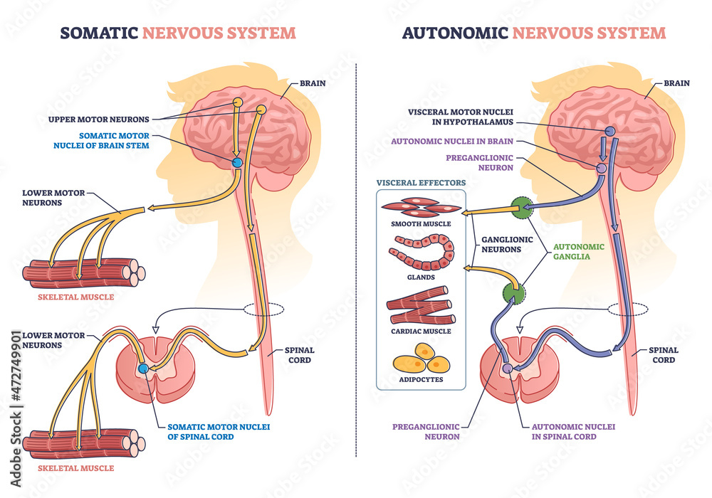
What Is a Neuron? Diagrams, Types, Function, and More - Healthline Sensory neurons are triggered by physical and chemical inputs from your environment. Sound, touch, heat, and light are physical inputs. Smell and taste are chemical inputs. For example, stepping on...
Action potential - Wikipedia An action potential occurs when the membrane potential of a specific cell location rapidly rises and falls. This depolarization then causes adjacent locations to similarly depolarize. Action potentials occur in several types of animal cells, called excitable cells, which include neurons, muscle cells, and in some plant cells.Certain endocrine cells such as pancreatic beta cells, and …
Glutamatergic synapses from the insular cortex to the ... - Neuron Apr 19, 2022 · Center for Neuron and Disease, Frontier Institute of Science and Technology, Xi’an Jiaotong University, Xi’an 710049, China ... and most of the FG + neurons were located within layer V, with far fewer FG-labeled neurons detected in the superficial layers ... Column diagram illustrating the PWMTs in different groups on day 14 (F (3,28) = 6. ...
Neuron Diagram Labeled Illustrations, Royalty-Free Vector Graphics ... Choose from Neuron Diagram Labeled stock illustrations from iStock. Find high-quality royalty-free vector images that you won't find anywhere else. Photos, Curated content, Curated sets, Signature collection, Essentials collection, Diversity and inclusion sets, Trending searches, Basketball game, Designer, Energy storage, Stressed,
Chloroplasts: Definition, Diagram, Structure and Function Apr 05, 2022 · Diagram of Chloroplast. The diagram of the chloroplast given below represents the chloroplast structure including the different parts of the chloroplast. The parts of a chloroplast such as the inner membrane, outer membrane, intermembrane space, thylakoid membrane, stroma, and lamella are all mentioned.
File:Complete neuron cell diagram en.svg - Wikipedia English: Complete neuron cell diagram. Neurons (also known as neurones and nerve cells) are electrically excitable cells in the nervous system that process and transmit information. In vertebrate animals, neurons are the core components of the brain, spinal cord and peripheral nerves. Own work.
A Labelled Diagram Of Neuron with Detailed Explanations - BYJUS A Labelled Diagram Of Neuron with Detailed Explanations, Biology, Biology Article, Diagram Of Neuron, Diagram Of Neuron, A neuron is a specialized cell, primarily involved in transmitting information through electrical and chemical signals. They are found in the brain, spinal cord and the peripheral nerves. A neuron is also known as the nerve cell.
Label Parts of a Neuron Diagram | Quizlet Label Parts of a Neuron Diagram | Quizlet, Label Parts of a Neuron, 4.1 (11 reviews) + −, Flashcards, Learn, Test, Match, Created by, cottonje, Terms in this set (14) Dendrites, receives impulses from other nerve cells, axon hillock, The cell body...the part of the cell that houses the nucleus and keeps the entire cell alive and functioning,
Describe the structure and function of neuron with labelled diagram ... Describe the structure and function of neuron with labelled diagram, Neurons are the fundamental unit of the nervous system specialized to transmit information to different parts of the body. Functions of a neuron, 1.Neurons are specialized cells of the nervous system that transmit signals throughout the body.
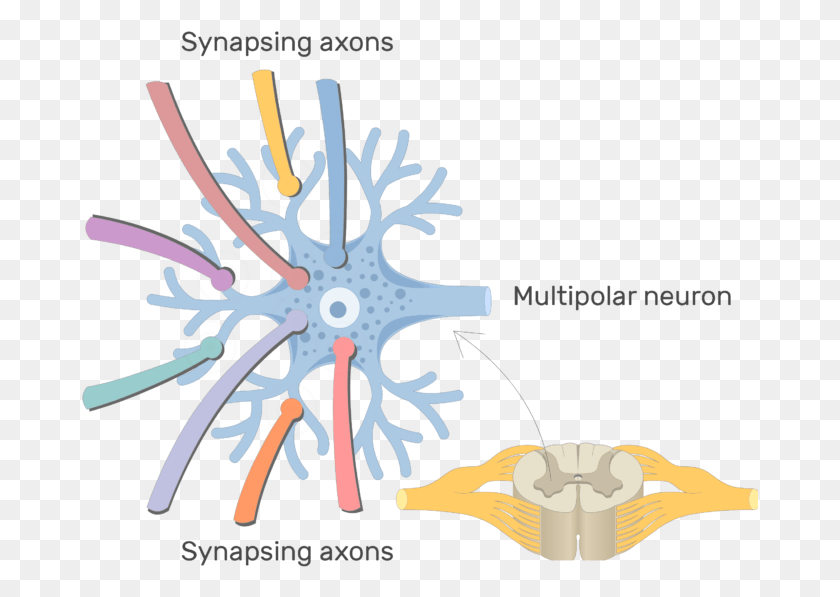
Nervous System – Medical Terminology for Healthcare Professions The major parts of the neuron are labeled on a multipolar neuron from the CNS. From Betts et al., 2013. Licensed under CC BY 4.0. ... This diagram shows a silhouette of a human highlighting the nervous system. The central nervous system is composed of the brain and spinal cord. The brain is a large mass of ridged and striated tissue within the ...
Diagram Quiz on Neuron Structure and Function (Labeling Quiz) This labelled diagram quiz on Neuron is designed to assess your basic knowledge in Structure and Function of Neuron. Choose the best answer from the four options given. When you've finished answering as many of the questions as you can, scroll down to the bottom of the page and check your answers by clicking ' Score'.
Label Neuron Anatomy Printout - EnchantedLearning.com Read the definitions, then label the neuron diagram below. axon - the long extension of a neuron that carries nerve impulses away from the body of the cell. cell body - the cell body of the neuron; it contains the nucleus (also called the soma) dendrites - the branching structure of a neuron that receives messages (attached to the cell body)
Neuron Diagram Labeled Pictures, Images and Stock Photos Search from Neuron Diagram Labeled stock photos, pictures and royalty-free images from iStock. Find high-quality stock photos that you won't find anywhere else. Video, Trending searches, Basketball, Fire, Texture, Chinese new year, Black history, Football, Popular categories, Lunar New Year Videos, Aerial videos, Abstract videos,














/purkinje_neuron-599da56d396e5a0011a0d344.jpg)
Post a Comment for "40 neuron diagram labeled"