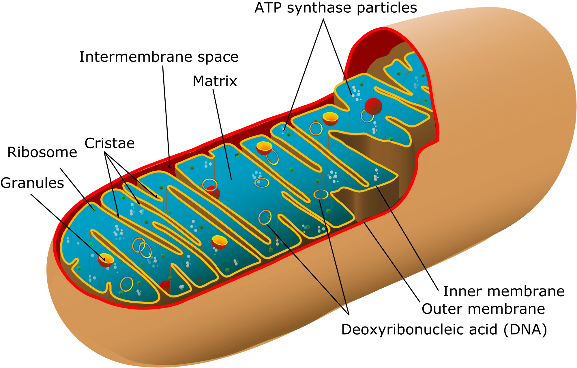
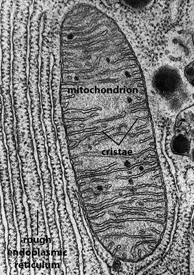
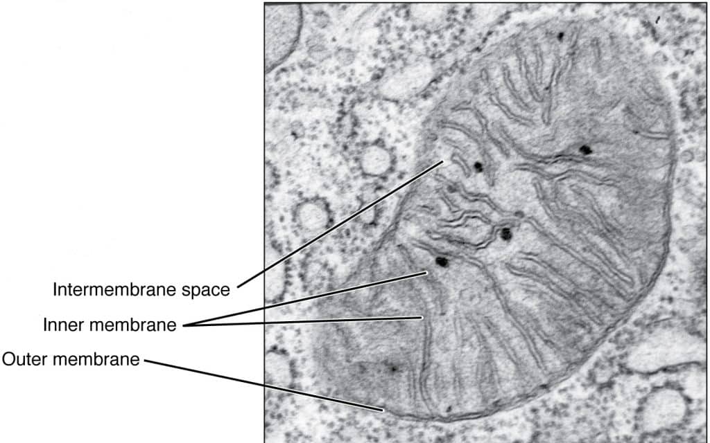
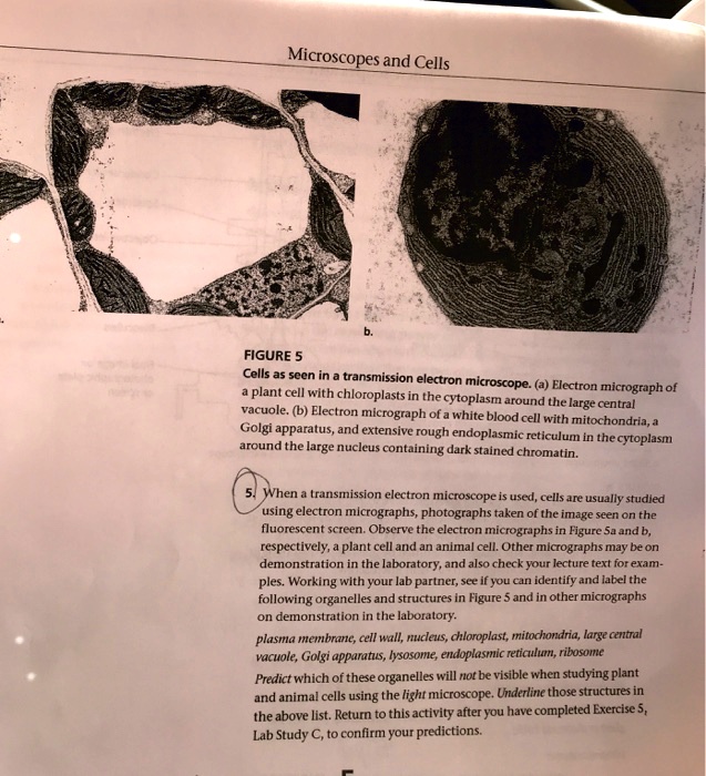
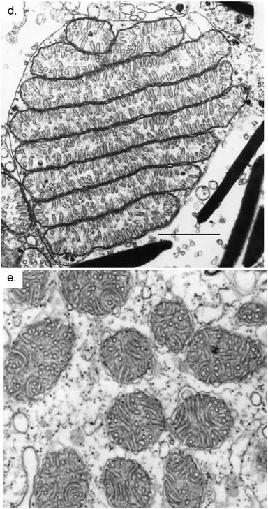
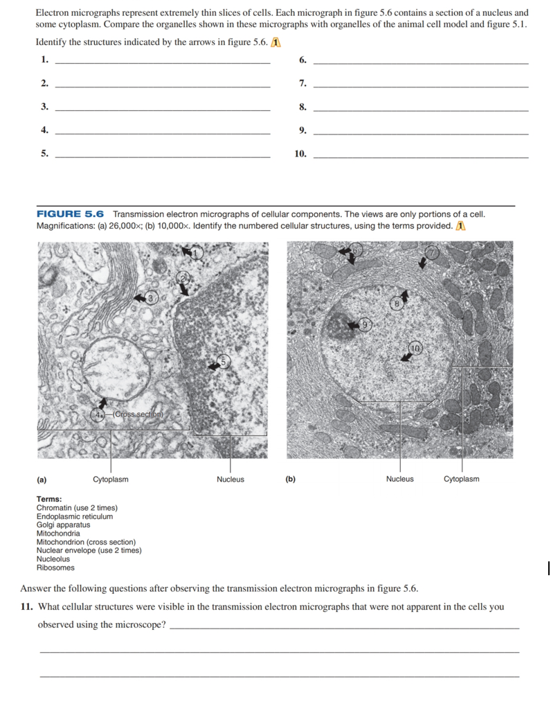
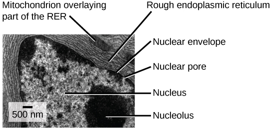
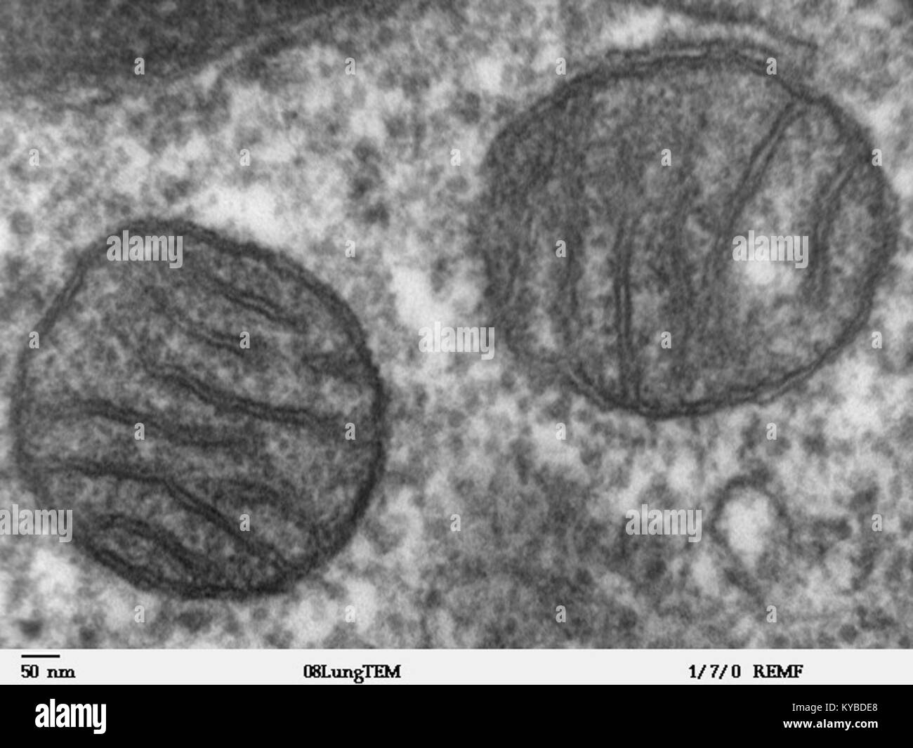
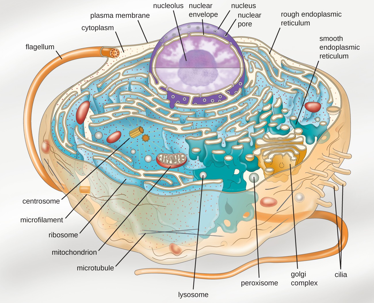
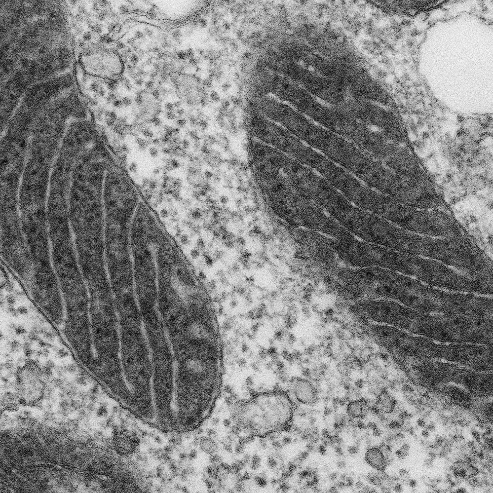
40 label the transmission electron micrograph of the mitochondrion
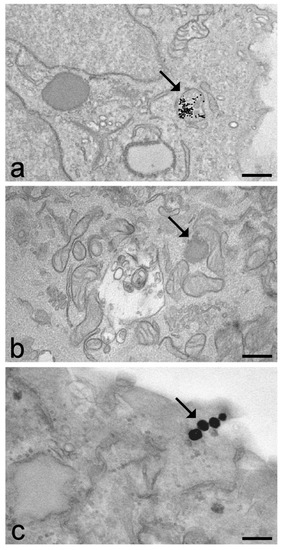
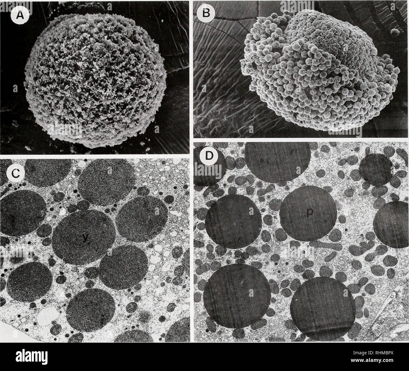
Transmission electron micrographs of mitochondria- nuage complexes in ... Download scientific diagram | Transmission electron micrographs of mitochondria- nuage complexes in spermatogonia of the sea urchin Anthocidaris crassispina. (A) Mitochondrion released the ... Draw the structure of a mitochondrion as seen in an electron micrograph ... 6)a) Draw the structure of a mitochondrion as seen in an electron micrograph. [5] B) Describe the central role of acetyl (ethanoyl) CoA in carbohydrate & fat metabolism. [5] Acetyl CoA is formed in both carbohydrate and fat metabolism. In carbohydrate metabolism, Acetyl CoA links glycolysis and the Krebs's cycle in a link reaction, in the link ...
quizlet.com › 651041260 › bio-hw-and-test-answersBio hw and test answers Flashcards | Quizlet c. Two atoms share one or more electron pairs unequally - polar covalent bone d. One example is water - polar covalent bond e. One example is a double bond between two carbon atoms - noncovalent bond f. Two atoms share one or more electron pairs roughly equally - noncovalent bond

Label the transmission electron micrograph of the mitochondrion
PDF Cambridge International Examinations Cambridge International Advanced ... 6 (a) Fig. 6.1 shows a transmission electron micrograph of a section through a mitochondrion. P Q Fig. 6.1 Table 6.1 shows some structures and compounds involved in aerobic respiration. Use letter P or letter Q from Fig. 6.1 to complete Table 6.1 to show the location of the structures and where the compounds are used. Table 6.1 › 34283602 › INTRODUCTION_TOINTRODUCTION TO BIOMEDICAL ENGINEERING - Academia.edu This article describes the experimental set-up and pharmacokinetic modeling of P-glycoprotein function in the rat blood-brain barrier using [11C]verapamil as the substrate and cyclosporin A as an inhibitor of P-gp. Transmission electron micrographs of mitochondria, site of ATP ... - Nature Transmission electron micrographs of mitochondria, site of ATP synthesis in eukaryotes All mitochondria retain core genomes of their own, which are necessary for the control of membrane...
Label the transmission electron micrograph of the mitochondrion. › 52500277 › Molecular_Cell_BiologyMolecular Cell Biology - Lodish - 5th Ed - Academia.edu Enter the email address you signed up with and we'll email you a reset link. Mitochondria electron micrograph - Big Chemical Encyclopedia Scanning electron micrograph of an H. rufescens spermatozoon. The sperm head, from mitochondrion (M) to tip of the acrosome vesicle (granule AV) is 7 pm. The width of the nucleus (N) is 1 pm. (Lower Panel). Transmission electron micrograph of the acrosomal vesicle showing it attached to the nucleus (NF) by the rod of actin filaments (AF). BIOL 230 Lecture Guide - Electron Micrograph of Mitochondria Transmission Electron Micrograph of a Mitochondrion Mitochondrion and surrounding cytoplasm of a cell from the pancreas of a bat. It is bounded by a smooth outer membrane about 7nm thick. An inner membrane has infoldings called cristae that project into the interior of the organelle. Rough endoplasmic reticula surround the mitochondrion. › 42749825 › Brock_Biology_ofBrock Biology of Microorganisms 13th Edition - Academia.edu Enter the email address you signed up with and we'll email you a reset link.
Solved Label the transmission electron micrograph of the - Chegg Label the transmission electron micrograph of the mitochondrion. Matrix granule Mitochondrion Outer membrane Cristae Inner membrane Matrix Reset Zoom Question: Label the transmission electron micrograph of the mitochondrion. Matrix granule Mitochondrion Outer membrane Cristae Inner membrane Matrix Reset Zoom This problem has been solved! How do you identify mitochondria in an electron micrograph? The mitochondria will appear circular if they are cut, in transverse section/across (the long axis). Describe the process of protein secretion/exporting. (It involves the rough ER, golgi apparatus, secretory vesicles, and the cell membrane.) The ribsomes attached to the rough ER produce proteins. Labeling the Cell Flashcards | Quizlet Label the transmission electron micrograph of the nucleus. membrane bound organelles golgi apparatus, mitochondrion, lysosome, peroxisome, rough endoplasmic reticulum nonmembrane bound organelles ribosomes, centrosome, proteasomes cytoskeleton includes microfilaments, intermediate filaments, microtubules Identify the highlighted structures Transmission electron micrograph showing the ultrastructure of a heart ... Heart mitochondria were isolated and fixed with 1% osmium tetraoxide and stained with uranyl acetate and lead citrate and examined with a Philips CM-10, Transmission Electron Microscope operated ...
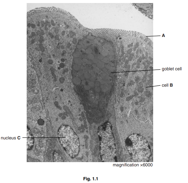
Solved Label the transmission electron micrograph of the - Chegg Transcribed image text: Label the transmission electron micrograph of the cell. 0 Nucleus rences Mitochondrion Heterochromatin Peroxisome Vesicle ULAR bumit Click and drag each label into the correct category to indicate whether it pertains to the cytoplasm or the plasma membrane. Transmission electron micrograph of mitochondria - Stock Image - G465 ... Mitochondria. Transmission electron micrograph of mitochondria from a mouse kidney cell. The large circle is a slice through a single mitochondrion. Note the double outer membrane and numerous internal membranes, or cristae, where respiration takes place. Mitochondria provide cells with energy by oxidising sugars and fats. Cells and magnification Flashcards | Quizlet 1- add a drop of water to slide 2- prepare a thin sample by using forceps to get some of the cells from the plant tissue 3- place them in a slide and again then with iodine solution 4- put on a cover slip and push down using a mounted needle The figure shows a microscopic image of a plant cell A Labelled Diagram Of Mitochondria with Detailed Explanation - BYJUS The term 'mitochondrion' is derived from a Greek word which refers to threadlike granules, and it was first described by German pathologist - Richard Altmann in 1890. Mitochondria are a double-membrane-bound cell organelle found in most eukaryotic organisms. In all living cells, these cell organelles are found freely floating within the ...
9+ label the transmission electron micrograph of the cell most standard ... 3.Solved Label the transmission electron micrograph of the | Chegg.com; 4.Electron Micrographs; 5.Cell Micrographs - BioNinja; 6.(A and B) Electron micrograph of a cell labeled for/5-tubulin followed… 7.anatomy 10.png - Label the transmission electron micrograph of the; 8.1.2 Skill: Interpretation of electron micrographs - YouTube
doku.pub › documents › nelson-biology-12pdf-30j71j2z320wNelson Biology 12.pdf [30j71j2z320w] - doku.pub The orbital that is occupied by an electron is what determines the energy level of the electron. The farther away the electron is from the nucleus, the greater its energy. The balloon-like 2p orbitals contain electrons that are farther away from the nucleus than the electrons in the 2s orbital, and thus hold the electrons with a higher energy ...
Transmission Electron Microscopy Study of Mitochondria in ... - PubMed Transmission Electron Microscopy Study of Mitochondria in Aging Brain Synapses Authors Vladyslava Rybka 1 , Yuichiro J Suzuki 2 , Alexander S Gavrish 3 , Vyacheslav A Dibrova 4 , Sergiy G Gychka 5 , Nataliia V Shults 6 Affiliations 1 Department of Pharmacology and Physiology, Georgetown University Medical Center, Washington, DC 20057, USA.
Transmission Electron Microscopy for Analysis of Mitochondria in Mouse ... Transmission Electron Microscopy for Analysis of Mitochondria in Mouse Skeletal Muscle Bio Protoc. 2018 May 20;8(10):e2455. doi: 10.21769/BioProtoc.2455. ... While a number of staining methods are available to study mitochondria, transmission electron microscopy (TEM) is still the most important method to study mitochondrial structure and ...
quizlet.com › 581840041 › lap-practical-1-ec-flash-cardsLap Practical #1 EC Flashcards | Quizlet Study with Quizlet and memorize flashcards containing terms like Place the following cytoplasmic structures in the appropriate structural category., Label the transmission electron micrograph of the cell., What would be the consequence if the highlighted structures suddenly became nonpolar? and more.
PDF Transmission Electron Microscopy Study of Mitochondria in Aging Brain ... mitochondria that may underlie declines in age-related synaptic function and may couple to age-dependent loss of synapses. Keywords: aging; brain; electron microscopy; hippocampus; mitochondria; synapse 1. Introduction Aging is a physiological, progressive, and time-dependent process that results in accumulated
The function of electron microscopy in contemporary surgical diagnostic ... The transmission electron microscope is comparable to a high-magnification, high-resolution light microscope in terms of the pathologist's ability to see minute intracellular and extracellular ...
› 41445571 › Ganong_s_Reviw_ofGanong’s Reviw of Medical Physiology 26th e. - Academia.edu information contained herein with other sources. For example and in particular, readers are advised to check the product information sheet included in the package of each drug they plan to administer to be certain that the information contained in this work is accurate and that changes have not been made in the recommended dose or in the contraindications for administration.
Transmission electron micrographs of mitochondria, site of ATP ... - Nature Transmission electron micrographs of mitochondria, site of ATP synthesis in eukaryotes All mitochondria retain core genomes of their own, which are necessary for the control of membrane...
› 34283602 › INTRODUCTION_TOINTRODUCTION TO BIOMEDICAL ENGINEERING - Academia.edu This article describes the experimental set-up and pharmacokinetic modeling of P-glycoprotein function in the rat blood-brain barrier using [11C]verapamil as the substrate and cyclosporin A as an inhibitor of P-gp.
PDF Cambridge International Examinations Cambridge International Advanced ... 6 (a) Fig. 6.1 shows a transmission electron micrograph of a section through a mitochondrion. P Q Fig. 6.1 Table 6.1 shows some structures and compounds involved in aerobic respiration. Use letter P or letter Q from Fig. 6.1 to complete Table 6.1 to show the location of the structures and where the compounds are used. Table 6.1











Post a Comment for "40 label the transmission electron micrograph of the mitochondrion"