39 sarcomere structure labeled
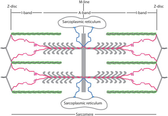
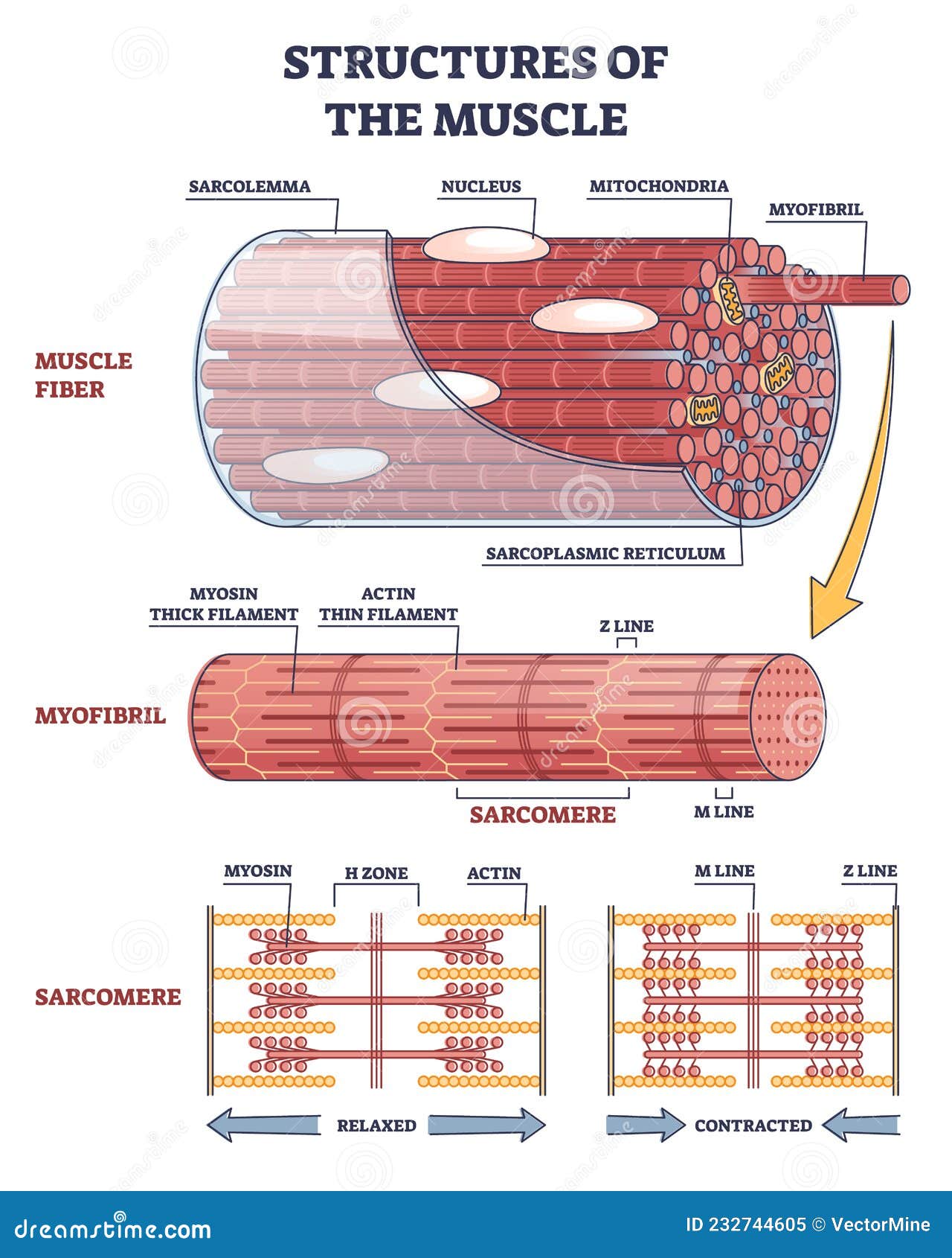
Diagram Of Sarcomere A sarcomere is the basic unit of striated muscle tissue. It is the repeating unit between two Z lines. Skeletal muscles are composed of tubular muscle cells which. sarcomere. Schematic: The Z line is depicted in black, myosin in red, actin in green/gray, and tropomyosin in blue. Image: MPI of Molecular Plant Physiology. Sarcomere definition. anatomy of a sarcomere contraction actin filament sliding muscle band diagram filaments zone skeletal anatomy myofilaments titin contract slide biology sarcomere myosin line relaxed. Depiction Of Muscle Sarcomere. Left: Micrograph Of A Sarcomere. Right. . sarcomere micrograph contracted depiction luzi depicting.
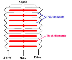
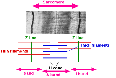
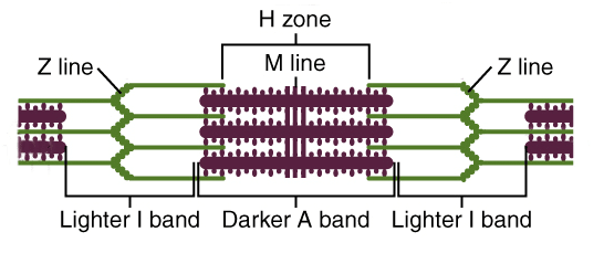
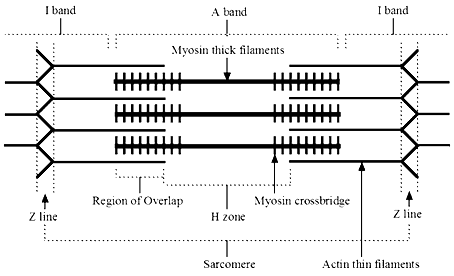
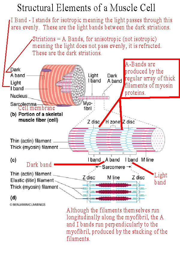
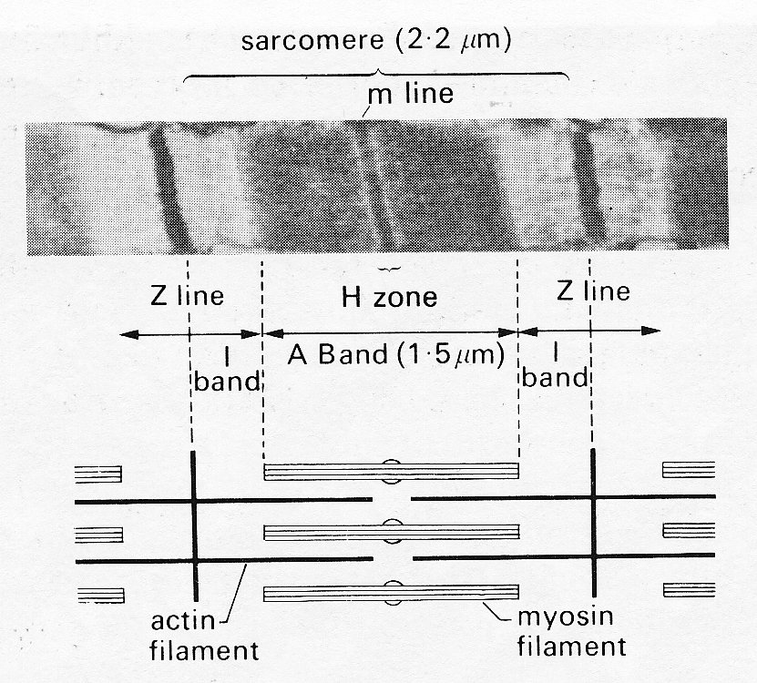
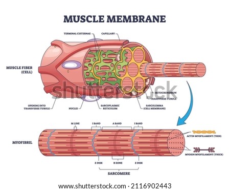
Sarcomere: Structure and Parts, Functions and Histology The main components of the histology of a sarcomere are summarized below: Band A Thick filament zone, composed of myosin proteins. Zone H Central zone of band A, without actin proteins superimposed when the muscle is relaxed. Band I Zone of thin filaments, composed of actin proteins (without myosin). Z disks
Sarcomere structure labeled
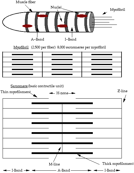
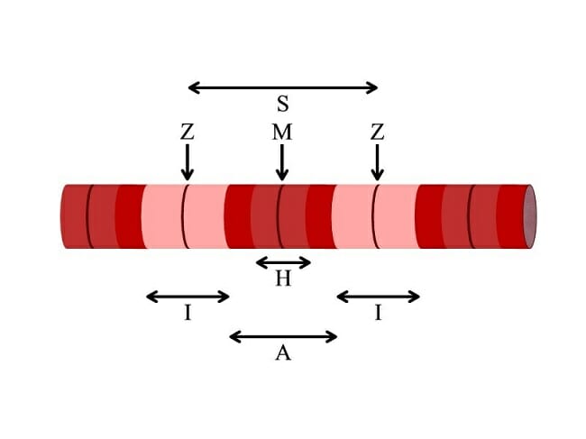
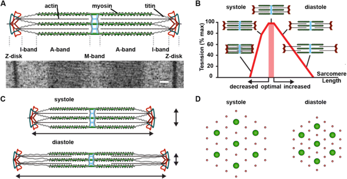
What is a Sarcomere? - Parts & Contraction - Study.com Each sarcomere has a central A-band which consists of thick filaments and two halves of I-band which has thin filaments. The I-bands from two neighboring sarcomeres meet at the Z-line, while the... Sarcomeres: "I" and "A" Bands, "M" and "Z" Lines, "H" Zone Each sarcomere divides into different lines, bands, and zone: "I" and "A" bands, "M" and "Z" lines, and the "H" zone. - Z-lines define the boundaries of each sarcomere. - The M-line runs down the center of the sarcomere, through the middle of the myosin filaments. - The I-band is the region containing only thin filaments. Labeled Sarcomere Diagram Labeled Sarcomere Diagram. Their observations led to the discovery of sarcomere zones. Sarcomere The figure depicts the structure of a Sarcomere. (Each zone is labeled). They first. Start studying Sarcomere Labeling. Learn vocabulary, terms, and more with flashcards, games, and other study tools. A sarcomere is the basic unit of striated muscle tissue.
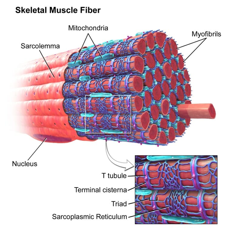
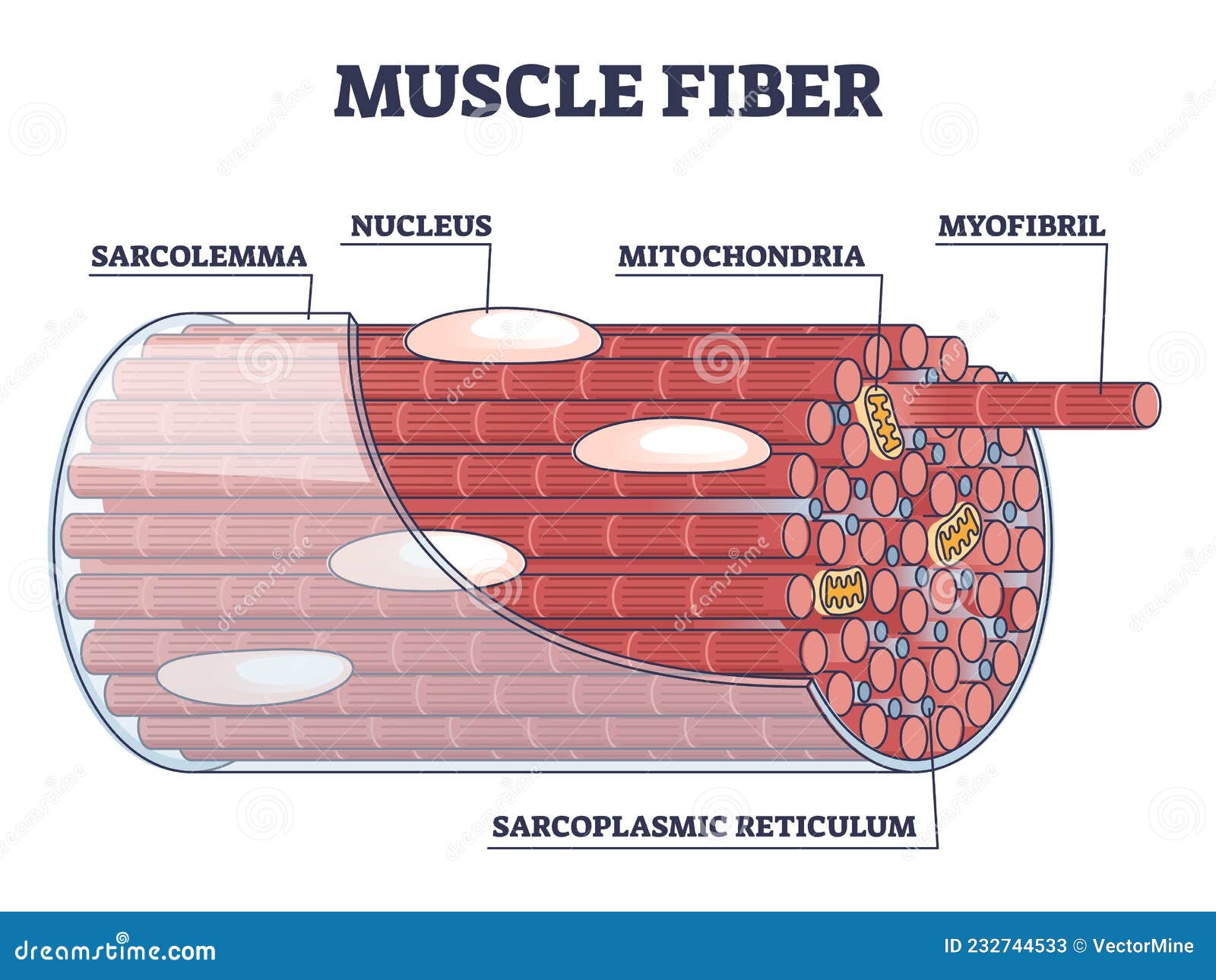
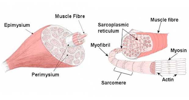
Sarcomere structure labeled. Label the Structures in a Sarcomere Quiz - purposegames.com This is an online quiz called Label the Structures in a Sarcomere. There is a printable worksheet available for download here so you can take the quiz with pen and paper. From the quiz author. Muscular System Quiz Practice This quiz has tags. Click on the tags below to find other quizzes on the same subject. Anatomy. Sarcomere (Muscle) Coloring - The Biology Corner The sarcoplasmic reticulum (E) is a network of tubes that run parallel to the myofilaments. Color this network green. The transverse tubules (C) run perpendicular to the filaments - color both yellow. The enter muscle fiber is surrounded by the sarcolemma (D), color this membrane brown. Label the Sarcomere Structure Diagram | Quizlet Label the Sarcomere Structure STUDY Learn Write Test PLAY Match Created by jack_burton76PLUS Terms in this set (12) z disc mysosin (thick) thin (actin) filament I band A band I band H zone elastic (titin) filaments elastic (titin) filaments thin (actin) filament thick (myosin) filaments myosin heads Sets found in the same folder Label the Sarcomere Sarcomere: anatomy, structure and function | Kenhub Each sarcomere is composed of protein filaments (myofilaments) that include mainly the thick filaments called myosin, and thin filaments called actin. The bundles of myofilaments are called myofibrils. The structure of the sarcomere is traditionally described with dark and light bands visible under the microscope. This banding pattern in sarcomeres is due mainly to the arrangement of thick and thin myofilaments in each unit.
human muscle structure labeled Sarcomere Model Sarcomere Structure - YouTube . sarcomere structure. PPT - Anatomy & Physiology H Jefferson Township High School Mrs . skull anterior human figure anatomy phyllis jefferson physiology township diagrams mrs smith ppt powerpoint presentation. HM Practical - Blood Vessel Histology - Embryology Sarcomere Labeling Quiz - PurposeGames.com This is an online quiz called Sarcomere Labeling. ... Label Parts of the Skull - Lateral View 10p Image Quiz. ... 13p Matching Game. Skeletal Muscle Matching - Back of the Body 10p Image Quiz. Integumentary System Labeling 7p Image Quiz. Muscle Structure Labeling 8p Image Quiz. Action Potential Graph 5p Image Quiz. Vertebral Column Labeling 7p ... Solved Label the skeletal muscle cell structures and - Chegg Expert Answer. 100% (8 ratings) Pls consider the boxes numbered from 1 to 10 I.e., the first box is 1 the second box is 2 and then the remaining as 3,4,5 till 10 1, The first box is SKELETAL MUSCLE FIBRE The skeletal muscles are together made up of many bundles called fasicles. Eac …. View the full answer. Sarcomere - Definition, Structure, Function and Quiz - Biology Dictionary Sarcomere definition. A sarcomere is the functional unit of striated muscle. This means it is the most basic unit that makes up our skeletal muscle. Skeletal muscle is the muscle type that initiates all of our voluntary movement. Herein lies the sarcomere's main purpose. Sarcomeres are able to initiate large, sweeping movement by contracting in unison.
Sarcomere - an overview | ScienceDirect Topics A sarcomere is the basic contractile unit of muscle fiber. Each sarcomere is composed of two main protein filaments—actin and myosin—which are the active structures responsible for muscular contraction. The most popular model that describes muscular contraction is called the sliding filament theory. In this theory, active force is generated ... Sarcomere | Definition, Structure, & Sliding Filament Theory - iBiologia Sarcomere Structure: Each sarcolemma or sarcomere is identical to biochemical composition to Plasmalemma, (that is another word for cell membrane). After observing under the microscope, a stacked pattern organized with a varied length of muscle fiber cells is seen. The bundle of filament arranged parallel to another is myofibril strands. Sarcomere Labeling Diagram | Quizlet Sarcomere The smallest contractile unit of muscle; extends from one Z disc to the next H Band The band at the middle of the A Band, where only myosin is found A Band The darkest area that runs the length of the myosin, including where actin and myosin overlap I Band On either side of the A Band is the I band, where only the Actin is found Z Disk Sarcomere Structure : Mnemonic | Epomedicine Z is the final alphabet: Z lines represents the end of sarcomere. M for middle: M line represents the midline of sarcomere. I is a thin letter: I band has only thin filaments. H is a thick letter: H zone has only thick filaments. A is a hybrid of "I" and "H": A band has both thin and thick filaments (remains constant during contraction)
Sarcomere: Structure and Parts, Functions and Histology The main components of the histology of a sarcomere are summarized below: Band A Thick filament zone, composed of myosin proteins. Zone H Central A-band zone, without overlapping actin proteins when muscle is relaxed. Band I Area of thin filaments, composed of actin proteins (without myosin). Z discs
Sarcomere - an overview | ScienceDirect Topics The sarcomere is the fundamental unit of contraction and is defined as the region between two Z-lines. Each sarcomere consists of a central A-band (thick filaments) and two halves of the I-band (thin filaments). The I-band from two adjacent sarcomeres meets at the Z-line.
Sarcomere Structure Tutorial | Sophia Learning | Human muscle anatomy ... Sarcomere Structure. I can identify the parts of a sarcomere. Samantha Meyers. ... ~* Anatomy *~ Science Projects. School Projects. A Level Biology. Homework Ideas. ... This is a tough unit to teach. Chemical structures, monomers, and polymers are so abstract! You will never teach biochemistry without these activities again! My students love my ...
muscle zones anatomy z zone Sarcomere labeled diagram structure muscle anatomy organization skeletal ppt powerpoint presentation 4b figure chapter within slideserve. Muscle anatomy & physiology muscle zones anatomy z zone. I. Identify The Main Muscles Of The Body, Using The Accompanying we have 9 Pictures about I. Identify The Main Muscles Of The Body, Using The ...
Solved 12. Label the following diagram of a sarcomere - Chegg Transcribed image text: 12. Label the following diagram of a sarcomere (filaments, bands, lines, etc., ...) ***Which regions shorten during contraction?
Sarcomere - Wikipedia A sarcomere (Greek σάρξ sarx "flesh", μέρος meros "part") is the smallest functional unit of striated muscle tissue. It is the repeating unit between two Z-lines. Skeletal muscles are composed of tubular muscle cells (called muscle fibers or myofibers) which are formed during embryonic myogenesis.Muscle fibers contain numerous tubular myofibrils.
Describe the structure of sarcomere. - Toppr Ask Sarcomeres are composed of long, fibrous proteins as filaments that slide past each other when a muscle contracts or relaxes. Two of the important proteins are:- 1) Myosin forms the thick filament. Myosin has a long fibrous tail and a globular head, which binds to actin. Its head also binds to ATP, which is the source of energy for muscle movement.
Sarcomere (Muscle) Coloring | Health science projects, Human muscular ... Myofilaments, mitochondria, annd tubules can all be identified and labeled on this image. Mar 12, 2015 - Learn the structure of a muscle fiber by coloring an individual sarcomere. Myofilaments, mitochondria, annd tubules can all be identified and labeled on this image. ...
Ultrastructure of Muscle - Skeletal - Sliding Filament - TeachMeAnatomy Ultrastructure of Muscle Cells. Muscle tissue has a unique histological appearance which enables it to carry out its function. There are three main types of muscle: Skeletal - striated muscle that is under voluntary control from the somatic nervous system. Identifying features are cylindrical cells and multiple peripheral nuclei.
To label: The given structure in the diagram of the sarcomere ... Textbook solution for Visual Essentials of Anatomy &Physiology 1st Edition Martini Chapter 6.1 Problem 2.3SR. We have step-by-step solutions for your textbooks written by Bartleby experts! To label: The given structure in the diagram of the sarcomere.
Sarcomere- Definition, Structure, Diagram, and Functions The structure of the sarcomere is described with the dark and the light band. A bands (or anisotropic bands): It is also called the dark band and contains the whole thick filament (myosin). I bands (or isotropic bands): it is called the light band that contains only the thin filament (actin).
Labeled Sarcomere Diagram Labeled Sarcomere Diagram. Their observations led to the discovery of sarcomere zones. Sarcomere The figure depicts the structure of a Sarcomere. (Each zone is labeled). They first. Start studying Sarcomere Labeling. Learn vocabulary, terms, and more with flashcards, games, and other study tools. A sarcomere is the basic unit of striated muscle tissue.
Sarcomeres: "I" and "A" Bands, "M" and "Z" Lines, "H" Zone Each sarcomere divides into different lines, bands, and zone: "I" and "A" bands, "M" and "Z" lines, and the "H" zone. - Z-lines define the boundaries of each sarcomere. - The M-line runs down the center of the sarcomere, through the middle of the myosin filaments. - The I-band is the region containing only thin filaments.
What is a Sarcomere? - Parts & Contraction - Study.com Each sarcomere has a central A-band which consists of thick filaments and two halves of I-band which has thin filaments. The I-bands from two neighboring sarcomeres meet at the Z-line, while the...
















Post a Comment for "39 sarcomere structure labeled"